Oropharynx (p16+)

Overview

HPV associated Oropharyngeal Carcinoma

  • HPV associated cancers of the oropharynx have a different biological behaviour than non-HPV associated cancers.
  • The disease more commonly affects younger and healthier patients with little to no tobacco exposure.
  • Prognosis for HPV related carcinomas is better than for HPV unrelated carcinomas.
  • HPV is classified as either low risk (causes benign neoplasms), or high risk (can cause malignancy).
  • HPV 16 and 18 are the most commonly detected oncologic strains (followed by HPV 31 and 33).
    • These two strains cause ≥ 70% of cervical cancer.
    • The majority of head and neck cancer is caused by HPV 16 (95%).
    • Overall, there are 15 high risk HPV subtypes (16, 18, 31, 33, 35, 39, 45, 51, 52, 56, 58, 59, 68, 73, and 82; from Robbins Basic Pathology).

HPV Infection in the Oropharynx

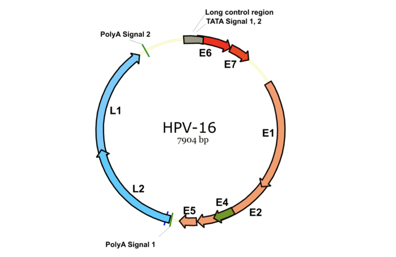
  • HPV is an encapsulated, non-enveloped dsDNA virus of the family papillomaviridae.
  • It has a circular viral DNA.
  • The virus is only productive and infectious in the basal cell layer (stratum basale) in squamous epithelium.
  • The basal cell layer is only exposed when there are microabrasions in the skin or when there are crypts.
    • The epithelia of the tonsils and base of tongue contain cryptic invaginations that expose the basal epithelial cells at their base, which predisposes them to HPV infection
    • This is very different from the oral cavity where there is a thick protective layer of stratified squamous mucosa.

Tumorigenesis with HPV Infection

  • Two of the proteins in the HPV genome inhibit tumour suppressors: E6 and E7.
    • The product proteins in the HPV genome are named by their temporal relationship to cell differentiation.
    • This is either early (E1-E7), or late (L1-L2).
    • E6 is an oncoprotein that inactivates p53
    • E7 is an oncoprotein that inactivates pRb
    • In HPV positive tumours, both wild type p53 and pRB are present but are inactivated.
  • During the initial phase of HPV infection, the viral DNA exists primarily in episomal form (not integrated into the host DNA)
  • If the virus is not cleared by the host, the viral DNA eventually integrates into the host genome, which then results in increased expression of E6 and E7.
  • Most infections are cleared rapidly by the host immune system, however in cases where the infection remains, there is a later risk of neoplasm.
  • Is is unclear why some individuals clear the virus while others do not.

p16 Overexpression as a Surrogate Marker for HPV

  • The p16INK4a (p16) tumour suppressor protein is over-expressed in HPV associated tumours, and is used as a surrogate to identify HPV associated carcinoma.
  • p16 is a protein that inhibits cyclin-dependent kinases 4/6 (CDK4/6)
    • Cyclin dependent kinases play a significant role in cell cycle progression.
    • In particular, CDK4 phosphorylates pRB which then triggers cell cycle progression.
    • Rb itself negatively regulates the production of p16 (negative feedback cycle).
    • As a result, inactivation of Rb (by viral E7) causes overexpression of p16

Changes from the AJCC 7th Edition

T-Categorization

  • Largely unchanged compared to the staging for p16- oropharyngeal carcinoma, with 2 exceptions:
    1. Carcinoma in situ (Tis) has been removed
    2. T0 is included (for cases where there is no clear primary but p16+ metastasis.
    3. T4b has been removed

N-Categorization

  • Now significantly different from nodal categorization for p16- oropharyngeal carcinoma.
  • Clinical categorization is based on the laterality and size of nodes.
  • Pathologic categorization is based on the number of nodes.
  • Extranodal extension is not a factor.

M-Categorization

  • Unchanged.

Group Staging

  • Group staging has been drastically changed.
  • Stage IV disease is reserved only for those with distal metastatic disease.

T-Categorization
T Category Criteria
T0

No primary tumour identified

T1

2 cm or smaller in greatest dimension.

T2

> 2 cm - 4 cm in greatest dimension

T3
  • > 4 cm in greatest dimension.
  • OR extension to the lingual surface of the epiglottis.
T4
  • Moderately advanced local disease
  • Tumour invasion of the larynx, extrinsic muscles of the tongue, medial pterygoid, hard palate, mandible, or beyond.
  • Tumour extension to the lingual surface of the epiglottis does not constitute invasion of the larynx (would be classified as T3 instead as above).

General Rules

  • Very similar to the T-categorization for p16- oropharyngeal carcinoma, with some exceptions:
    1. Any advanced local disease is categorized as T4 (there is no T4b categorization unlike with p16- oropharyngeal carcinoma).
    2. T0 is considered a category (in situations where there is p16+ metastasis but no clear primary).
    3. Tis has been removed.

N-Categorization

Clinical Nodal Categorization

N Category Criteria
NX

Regional lymph nodes cannot be assessed.

N0

No regional lymph node metastasis.

N1

One or more ipsilateral lymph nodes, all measuring 6 cm or less

N2

Bilateral or contralateral lymph nodes, all measuring 6 cm or less

N3

Nodal metastasis with any lymph node measuring > 6 cm

Pathologic Nodal Categorization

N Category Criteria
NX

Regional lymph nodes cannot be assessed.

N0

No regional lymph node metastasis.

N1

Metastasis in 4 or fewer lymph nodes

N2

Metastasis in more than 4 lymph nodes

Notes

  • Clinical nodal categorization includes an N3, but pathologic nodal categorization excludes N3
  • This decision was based on data from the Washington University School of Medicine, which found within their surgically managed dataset that N3 disease behaves unusually well and is equivalent to N1.
  • Their data showed that high metastatic node number rather than extracapsular spread, laterality, or nodal size is the key prognosticator in surgically resected, neck dissected p16+ oropharynx cancer.
  • ENE did not seem to have the same prognostic significance as with p16 negative disease.

M-Categorization
M Category Criteria
M0

No distant metastasis

M1

Distant metastasis present


Group Staging

Clinical Group Staging

N0
M0
N1
M0
N2
M0
N3
M0
Any N
M1
T0 I II III IV
T1 I I II III IV
T2 I I II III IV
T3 II II II III IV
T4 III III III III IV

Pathologic Group Staging

N0
M0
N1
M0
N2
M0
Any N
M1
T0 I II IV
T1 I I II IV
T2 I I II IV
T3 II II III IV
T4 II II III IV

General Rules

  • Clinical staging is more commonly used as the majority of oropharyngeal cancers are currently treated with radiotherapy +/- chemotherapy as the primary modality.
    • For this, only metastatic disease upstages you to Stage IV
    • Otherwise, if you have a T4 or N3 categorization, you are staged as a Stage III
    • Stage II and Stage I can be visualized by working upwards and leftwards on the table above.